Fluorescent polymer nanoparticles


Download the full application note here
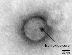
These fluorescent polymer nanoparticles are designed as molecular bioimaging probes with a core-shell structure, manufactured by StreamBio. A light emitting polymer is wrapped around an iron oxide core, then coated with a water-friendly capping agent. Using transmission electron microscopy, we can see the size and shape of the nanoparticles, as well as the location of the metal core inside them.
Negative staining is used to highlight the polymer nanoparticles, which would otherwise have too low contrast to be visible. We also use this technique to image many other organic materials, such as proteins, viruses and hydrogel fibres.
To make sure all the nanoparticles fluoresce in the same way, the size distribution needs to be small. Various techniques exist for measuring the size of nanoparticles, but they only give averages, which miss a lot of important information. TEM is the perfect tool for the job, allowing us to directly image individual nanoparticles. This gives us a wealth of information about size, shape and uniformity.

